Head: AMAGASA Toshiyuki, Professor, Ph.D.
Objectives
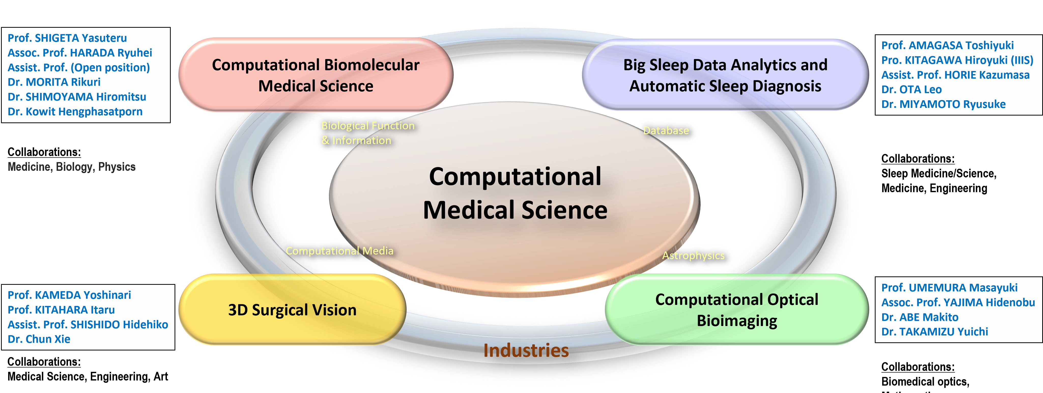
For the development of novel technologies for medical care, there has been hitherto conducted ”medicine and engineering cooperation”, which is the cooperation between medical scientists in private enterprises and engineers in academic institutes, or ”medicine, science and engineering cooperation” including natural scientists as well. Owing to recent rapid strides in computational science, state-of-the-art computational techniques have been developed, and they have established an indispensable research method comparable to theory and experiments. Besides, the technology of big data and machine learning has made remarkable progress in the fields of database and computational media. In Center for Computational Sciences, University of Tsukuba, we promote ”Computational Medical Science”, which initializes a novel approach of “medicine and computation cooperation” by incorporating cutting-edge computational sciences into medical science. In this department, we explore medical technologies based on the latest development of computational methods, imaging technology, and machine/deep learning, in cooperation of computational sciences in physics, life science, database technology, computational media engineering with medical science and industries. For the objectives, we propel four projects of (1) Computational Biomolecular Medical Science, (2) Big Sleep Data Analytics and Automatic Sleep Diagnosis, (3) 3D Surgical Vision, and (4) Computational Optical Bioimaging, in pursuing mutual cooperation and synergy.
Project teams
Computational Biomolecular Medical Science
Members:
Prof. SHIGETA Yasuteru
Assoc. Prof. HARADA Ryuhei
Assist. Prof. (Open position)
Dr. MORITA Rikuri
Dr. SHIMOYAMA Hiromitsu
Dr. Kowit Hengphasatporn
Collaborations:
Medicine, Biology, Physics
Overview:
In conventional drug development, one has aimed to develop small molecular drugs with low side effects that have both high selectivity and the binding ability for a specific target. In order to support them, in silico technology has been advanced in recent years, with the expectation that it will be possible to reduce the cost to 100 billion yen per drug for ten years. However, despite many attempts, few cases have been put to practical use because of the limitation of the small-sized drugs. One of the new drug targets is the middle-sized drugs such as cyclic polypeptides and DNA/RNA aptamers. In this group, we conduct researches on reaction pathway analysis using first-principles calculation and analysis of substrate-protein binding process using molecular dynamics calculation for drug discovery targeting on middle-sized molecules. In particular, the computational methodologies for the membrane permeability, pKa, and redox potential prediction are developed.
Big Sleep Data Analytics and Automatic Sleep Diagnosis
Members:
Prof. AMAGASA Toshiyuki
Prof. KITAGAWA Hiroyuki (IIIS)
Assist. Prof. HORIE Kazumasa
Dr. OTA Leo
Dr. MIYAMOTO Ryusuke
Collaborations:
Sleep Medicine/Science, Medicine, Engineering
Overview:
Sleep plays an important role in people’s daily lives. Sleep disorders could cause various illnesses. There are a lot of scientific questions that need to be answered about sleep. Quantitative and accurate measurements and analysis of sleep are fundamental issues in sleep research. Sleep polysomnography (PSG) is a commonly used sleep measurement method. PSG attaches many sensors to the subject and continuously measures and records various biological data such as brain waves, respiratory movements, and eye movements throughout the night. In PSG, the burden and cost of the subject are large, and measurement over a long period is impossible. Moreover, analysis of the acquired data depends on manual inspection by human experts, and it is impossible to analyze large-scale data. In response to the increasing social interest in sleep in recent years, some methods have been developed to easily measure sleep status using a smartphone or the like. However, at present, there is still no means to conveniently measure sleep as accurately as human experts. This research aims to realize automatic sleep analysis and diagnosis by integrating sleep big data analysis, machine learning and new sensing technology, and to pioneer a new computational medical science field.
3D Surgical Vision
Members:
Prof. KAMEDA Yoshinari
Prof. KITAHARA Itaru
Assist. Prof. SHISHIDO Hidehiko
Dr. Chun XIE
Collaborations:
Medical Science, Engineering, Art
Overview:
Although the safety of surgery has improved urgently due to advances in medical technology, the upper limit of patient survival is always surgical cancer, the number one cause of death in Japan, and the 1st treatment option is surgery. On the other hand, doctors vary in skills because of increased knowledge and skills that need to be acquired and lost due to medical progress (regional disparities in 5-year survival rates for cancer resection are more than 10%). Since education takes about 15 years, there is a shortage of surgeons and there are concerns that the problem will become even more serious. Until now, research and development on virtual surgery using IT technology has been carried out, however there are not many implementation examples of surgical navigation systems based on 3D visual information. Our research aims to realize “3D surgical navigation system” that integrates “3DCG simulation” used by doctors for preoperative examination and “on-site navigation” based on 3D Computer Vision technology. Supporting surgeons with an intuitive understanding of proper instructional procedures and advanced medical techniques can improve the level of surgery. In addition, by utilizing technologies such as 3DCG modeling and Augmented Reality (AR), we realize a remote diagnosis and VR treatment system, which aim to correct regional disparities in medical technology. Furthermore, by applying our technologies to laparoscopic surgery, a surgical procedure with less physical burden on the patient can be possible.
Computational Optical Bioimaging
Members:
Prof. UMEMURA Masayuki
Assoc. Prof. YAJIMA Hidenobu
Dr. ABE Makito
Dr. TAKAMIZU Yuichi
Collaborations:
Biomedical optics, Mathematics
Overview:
Recent years, the technology of optical imaging is extraordinarily developing, and thereby molecular processes in live cells and tissues can be pursued. At present, the imaging methods such as X-ray CT or MRI are widely applied, and mainly detect the alteration of morphology at a level of organs and tissues. However, these methods are subject to the risk of significant exposure and require a large-scale apparatus or a specialized measurement room. On the other hand, the optical imaging can dynamically visualize multi-level function and morphological alteration in molecules and organs, and possesses a lot of advantages such as no exposure, non-invasion, bed side inspection, low cost, low restraint, and high time –resolution. Thus, it allows us to make automatic and remote diagnosis, monitoring, and screening and is applicable to new-born babies and infants without side effects like CT or MRI. Since biological tissues are strong scattering media, a sophisticated technology of computational science is required to extract the local information from scattered light. The study for the practical realization in clinical settings has not been thitherto sufficient. In this project, we aim at developing a safe technology of CT with near-infrared imaging under the cooperation among the construction of light propagation models based on radiative transfer equation (RTE), the inverse-problem analysis based on mathematical methodology, and the experiments of medical optics for biological tissues.
(Update: Apr. 1, 2022)